Since the development of this group, we have been exploring the development of instrumental methodology based on the accumulation of in situ spectroscopy electrochemical instruments developed in the previous period. In the process of research, on the basis of improving/optimizing the existing commercial instruments, the group has built its own instruments to adapt to the current environmental conditions and meet the current experimental needs, and has continuously formed a complete research system, which has become a new and efficient instrumentation methodology. At the same time, based on teaching, the group has developed a variety of teaching instruments suitable for undergraduate/postgraduate experimental teaching, which have been put into the first-line teaching practice and have been well received by teachers and students.The developed instruments are as follows:
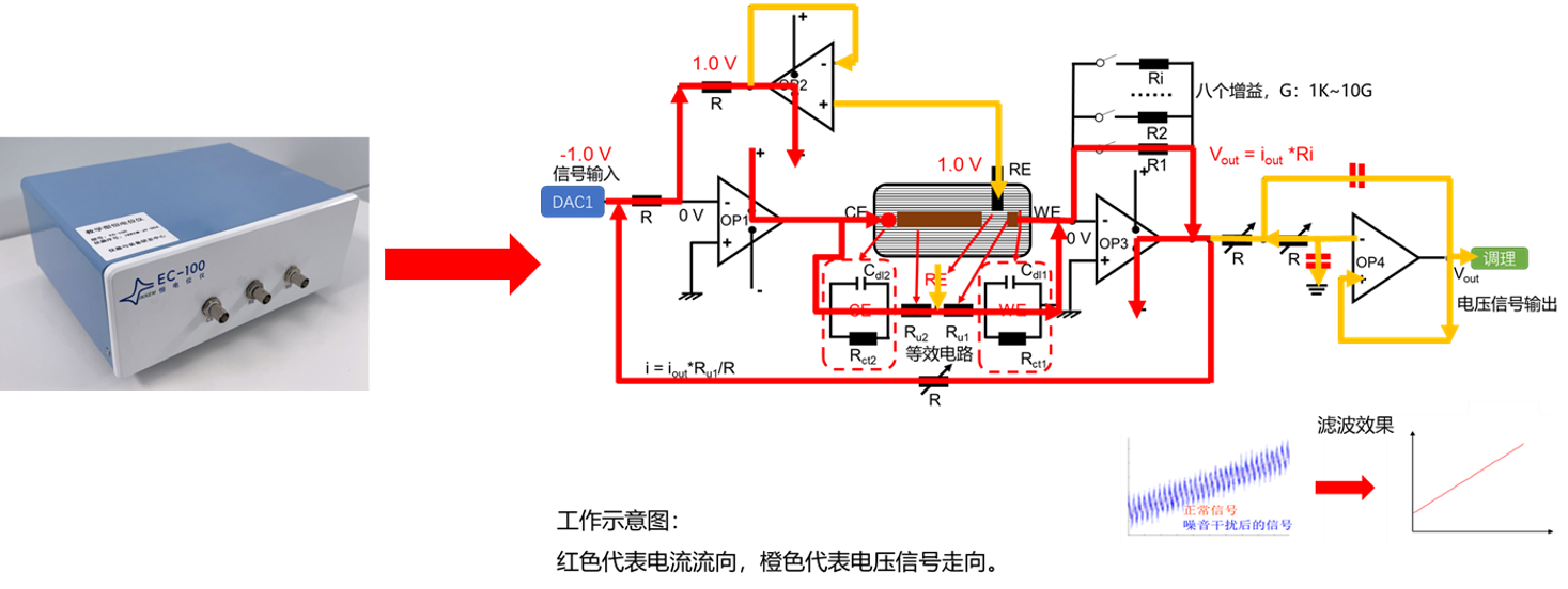
1. The potentiostat for electrochemical teaching experiments
Compared to other potentiostats available in the market, it boasts a user-friendly operation interface, excellent safety performance, and data recording and analysis capabilities. These features facilitate students' deeper comprehension of electrochemical principles and enhance the effectiveness of their experimental research. To date, the teaching potentiostat has been delivered, and the transition from a teaching to a research potentiostat is ongoing.
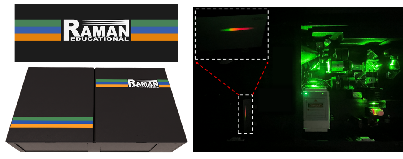
2. Educational Multifunctional Raman Spectrometer
Enabling sensitive detection and hands-on learning for in-class and research applications, adaptable for both solid and liquid sample analysis.
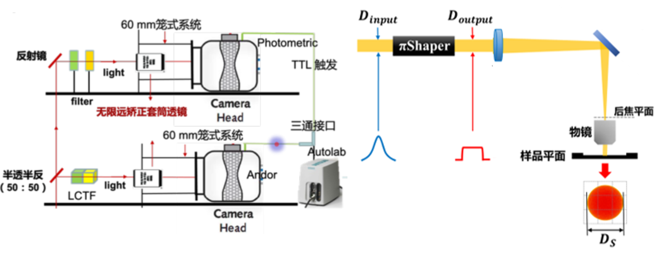
3. Home-made wide-field Raman imaging system
Developing multiplexing and flat-top illumination wide-field Raman imaging methods with high time resolution and spatiotemporal synchronization for understanding the electrochemical interface structure and reaction kinetics.
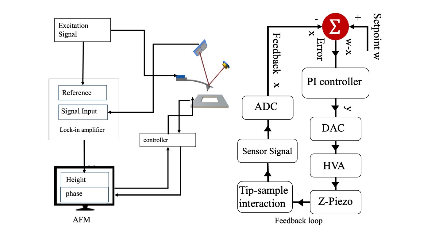
4. Ongoing research:AFM (Atomic Force Microscopy): A high-resolution atomic force microscopy technique that uses a probe to scan the surface of a sample and measure the interaction forces between atoms, thereby obtaining information on the morphology and physical properties of the sample surface. As the tip scans the surface, it experiences various forces, such as van der Waals forces, electrostatic forces, and magnetic forces. These forces cause the cantilever to bend, and this bending is detected by a laser beam reflected off the back of the cantilever. The deflection of the laser beam is then used to create a topographic map of the sample surface.
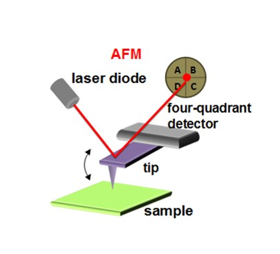
Development and construction of AFM for TERS. This AFM needs to be able to operate in an amplitude modulation mode. While imaging, the distance between the tip and sample is controlled to obtain a near-field Raman signal for TERS mapping.
5. Based on the three generations of EC-TERS that have been developed so far, we are innovatively carrying out the research and development of EC-TERS 4.0.

